Truffle Genus: Hydnangium
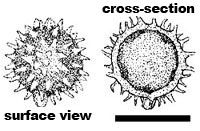 Hydnangium carneum basidiospore scale = 10 µm |
Kingdom: Fungi Phylum: Basidiomycota Order: Agaricales Family: Hydnangiaceae |
Spore Characters
Surface: Ornamented with hyaline spines and narrow cones, 1.5-2.5 µm tall and up to 2 µm broad at the base.
Shape and Size: Globose to subglobose, 10-18 µm in diameter, sterigmal attachment straight, inconspicuous.
Wall: Single, 1-2 µm thick.
Color in Water: Hyaline.
Melzer's Reaction: Not distinctive.
Comments: Hydnangium carneum is closely related to the mushroom genus Laccaria in the family Tricholomataceae. The two genera cannot be differentiated on the basis of spores alone.
View photos of Hydnangium spores
Sporocarp Characters
Shape and Size: Subglobose to turbinate, generally with a basal protuberance, 0.5-3 cm broad.
Peridium: Pale pink to pink or rose when fresh, fading to white with loss of moisture, felty; rhizomorphs lacking.
Gleba: Small to labyrinthine chambers separated by pink to rose chamber walls; base with a pad or projection of pink tissue that sometimes extends into the gleba as a simple to branched columella.
Odor: Not distinctive.
View photos of Hydnangium sporocarps